Fascination About Remote Patient Monitoring Devices |
Table of ContentsAn Unbiased View of Blood Pressure Monitor WatchRespiration Rate Monitoring Can Be Fun For AnyoneSome Known Facts About Body Temperature Watch.Unknown Facts About Wearable Ecg MonitorOxygen Reader Can Be Fun For AnyoneThe Facts About Blood Oxygen Monitor RevealedHow Remote Patient Monitoring can Save You Time, Stress, and Money.Getting My Measure Blood Pressure At Home To WorkLittle Known Facts About Home Sleep Apnea Test.The Single Strategy To Use For Medical Heart Rate MonitorA Biased View of Portable Ecg MachineThe Ultimate Guide To Respiration Monitor
Electrocardiogram depicting ventricular fibrillation in an individual with a left ventricular assist tool (LVAD). Ventricular fibrillation is frequently as a result of heart disease and can result in myocardial infarction and/or abrupt fatality. The rhythm on this electrocardiogram (ECG) is sinus with borderline PR prolongation. There is evidence of an acute/evolving anterior ischemia/myocardial infarction (MI) superimposed on the left bundle branch blocklike (LBBB) pattern.
Although this finding is not especially sensitive for ischemia/MI with LBBB, such main T wave modifications are fairly particular. The famous voltage with left atrial problem as well as leftward axis jointly with the left ventricular intraventricular transmission hold-up (IVCD) follow underlying left ventricular hypertrophy. This ECG is an instance of "bundle branch block plus." Picture politeness of http://ecg.
edu. This electrocardiogram (ECG) reveals proof of severe left ventricular hypertrophy (LVH) with prominent precordial voltage, left atrial irregularity, side ST-T irregularities, and a rather leftward QRS axis (15 ). The individual had malignant hypertension with intense cardiac arrest, bookkeeping additionally for the sinus tachycardia (blood stress at first 280/180 mmHg).
However, the ECG is not constant with considerable inferolateral coronary infarction. Photo thanks to http://ecg. bidmc.harvard. edu. The rhythm on this electrocardiogram is atrial tachycardia (price, 154 beats/min) with a 2:1 atrioventricular (AV) block. Keep in mind the partially hidden, nonconducted P waves on the ST sectors (eg, leads I as well as aVL).
The rSR' type complex in the side leads (I, aVL) is not due to an appropriate bundle branch block (RBBB) but to an atypical left ventricular transmission flaw. These unanticipated rSR' complexes in the side leads (El-Sherif indication) correlate with underlying considerable heart attack (MI) and also, sometimes, ventricular aneurysm. (El-Sherif.
1970; 32:440 -8.) The notching on the upstroke of the S waves in lead V4 with a left bundle branch block-type pattern also suggests underlying MI (Cabrera indicator). This patient had extreme cardiomyopathy second to coronary artery condition, with extensive left ventricular wall motion abnormalities. Image courtesy of http://ecg. bidmc.harvard. edu.
Such artifact may be created by a range of aspects, including inadequate electrode contact, muscle tremor, and electrical interference. A single premature ventricular facility (PVC) is existing with a countervailing time out such that the RR period bordering the PVC is twice as lengthy as the coming before sinus RR interval. Evidence of a previous anterior heart attack exists with pathologic Q waves in leads V1-V3.
Heart catheterization showed a 90% constriction in the person's proximal section the left former coming down coronary artery, which was treated with angioplasty as well as stenting. Broad P waves in lead V1 with a prominent unfavorable element follows a left atrial problem. Image thanks to http://ecg. bidmc.harvard. edu. This electrocardiogram (ECG) is from a person that went through immediate cardiac catheterization, which disclosed diffuse severe coronary convulsion (most marked in the left circumflex system) without any kind of set obstructive sores.
A question of supposed takotsubo cardiomyopathy (left ventricular apical ballooning syndrome) is additionally increased (see Bybee et al. Systematic review: short-term left ventricular apical ballooning: a syndrome that resembles ST-segment elevation myocardial infarction. Ann Int Medication 2004:141:858 -65). The latter is most usually reported in postmenopausal, middle-aged to senior women in the context of acute psychological anxiety as well as may trigger ST altitudes really with succeeding T wave inversions.
Myocarditis may additionally be associated with this kind of ECG as well as the cardiomyopathic searchings for revealed right here. No repaired obstructive epicardial coronary sores were discovered by coronary arteriography. The searchings for in this ECG consist of low-amplitude QRS facilities in the limb leads (with an indeterminate QRS axis), loss of typical precordial R wave progression (leads V1-V3), and also prominent anterior/lateral T wave inversions.
bidmc.harvard. edu. This electrocardiogram reveals a substantial acute/evolving anterolateral myocardial infarction pattern, with ST-T changes most noticeable in leads V2-V6, I, and aVL. Slow-moving R wave development is also existing in leads V1-V3. The rhythm is borderline sinus tachycardia with a single premature atrial complex (Political Action Committee) (4th beat). Note also the reduced arm or leg lead voltage as well as likely left atrial problem.
Picture thanks to http://ecg. bidmc.harvard. edu. This electrocardiogram shows a person is having a progressing anteroseptal myocardial infarction additional to drug. There are Q waves in leads V2-V3 with ST sector altitude in leads V2-V5 related to T-wave inversion. Additionally kept in mind are biphasic T-waves in the substandard leads. These multiple problems recommend occlusion of a huge left anterior coming down artery that twists around the apex of the heart (or multivessel coronary artery condition).
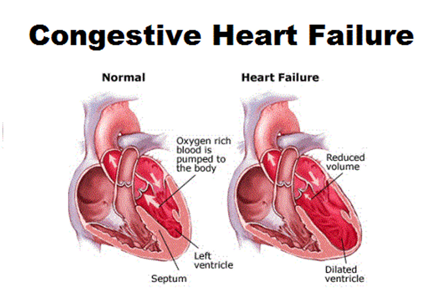
bidmc.harvard. edu. A color-enhanced angiogram of the heart left shows a plaque-induced blockage (leading center) in a major artery, which can cause heart attack (MI). MIs can speed up heart failing. Emphysema is consisted of in the differential diagnosis of heart failing. In this radiograph, emphysema bubbles are kept in mind in the left lung; these can severely hinder breathing capacity.
In this color Doppler and spooky Doppler ultrasonographic examination of the left internal carotid artery (ICA) in an https://www.washingtonpost.com/newssearch/?query=Heart health individual with cervicocephalic FMD, stenoses of around 70% is seen in the ICA. Cervicocephalic fibromuscular dysplasia (FMD) can bring about difficulties such as hypertension and chronic kidney failing, which, consequently, can bring about cardiac arrest.
Electrocardiogram from a 46-year-old man with enduring high blood pressure revealing left atrial irregularity and also left ventricular hypertrophy with stress. Electrocardiogram from a 46-year-old guy with enduring hypertension revealing left atrial problem as well as left ventricular hypertrophy with strain. Apical 4-chamber echocardiogram in a 37-year-old male with arrhythmogenic appropriate ventricular dysplasia (ARVD), a genetic cardiomyopathy.
ARVD can lead to ventricular and supraventricular arrhythmias. The most considerable of all rhythms connected with cardiac arrest are the life-threatening ventricular arrhythmias. Cardiac magnetic resonance image (CMRI), brief axis sight. This image shows best ventricular dilatation, trabucular derangement, aneurysm development as well as dyskinetic cost-free wall in an individual with arrhythmogenic right ventricular dysplasia.
Echocardiogram of a person with severe pulmonic stenosis. This photo reveals a parasternal brief https://www.cardiacsense.com/ axis view of the thickened pulmonary valve. Pulmonic constriction can result in pulmonary high blood pressure, which can result in hepatic blockage as well as in right-sided heart failing. Echocardiogram of a person with serious pulmonic stenosis. This photo reveals a Doppler scan of the height speed (5.
Echocardiogram of an individual with extreme pulmonic stenosis. This photo shows that reasonably severe pulmonary lack (orange color circulation) is also existing. This video clip is an echocardiogram of a patient with extreme pulmonic constriction. The initial section reveals the parasternal short axis view of the thickened lung valve. The second sector shows the existence of moderate lung deficiency (orange color flow).
Transesophageal echocardiogram with constant wave Doppler interrogation across the mitral shutoff showing an enhanced mean slope of 16 mm Hg regular with severe mitral stenosis.
This is the prominent heart problem organization in the United States. Their online resources on heart failure deal clear explanations of the condition for both clients and relative, in addition to links to numerous various other devices as well as resources. You can likewise discover details on your local AHA workplace and also discover volunteer possibilities.
It likewise has beneficial details for individuals coping with a heart condition in their person section. You can find out realities about cardiac arrest, and find out about applications as well as podcasts to aid you much better handle the condition. The American University of Cardiology's web site has plenty of resources for patient education and learning as well as empowerment.
By creating a profile, you can register to get tailored e-newsletters and also discussion overviews to bring with you to your following doctor's consultation. This tool from the American Heart Organization can be used on a smart device or desktop to track every little thing from everyday exercise to medications. You can also set it approximately advise you when it's time for your everyday doses.
 Some Ideas on Heart Failure Management You Should Know
This app can aid you to track your fluid consumption to prevent excess fluid accumulation, which can bring about difficulties. You can also track your weight, given that sudden rises in weight can be because of water retention. The wonderful point regarding this app is that it alerts you if your blood pressure or weight obtains high sufficient that you should tell your medical professional.
| Комментировать | « Пред. запись — К дневнику — След. запись » | Страницы: [1] [Новые] |


